
BM(Hons) MD FRCS(Neurosurgery)
Consultant Neurosurgeon
BrainTumourSurgery.co.uk
The most common investigations to undergo for a suspected brain tumour are a Computed Tomography (CT) scan and/or a Magnetic Resonance Imaging (MRI) scan. They may be performed for initial investigation or to help in the planning of your surgery (for neuronavigation), in which case you may have some small plastic dots (fiducials) stuck onto your head before the scan.
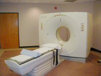
CT is an imaging test in which many x-rays are taken of the head from different angles. These images are combined by a computer to produce cross-sectional pictures of the brain. Except for the injection of a dye (needed in most but not all cases), this is a painless procedure that can be done as an outpatient. Modern CT scans take only a few minutes to perform and involve lying on a table that moves you into the scanner (which is open at both ends), and staying still for a few minutes.
As a CT uses X-rays (which may harm a foetus), it is important to know if female patients are pregnant so that the abdomen can be shielded or alternative scanning techniques used. Any known allergy to contrast (dye) should be reported.